


Product Description
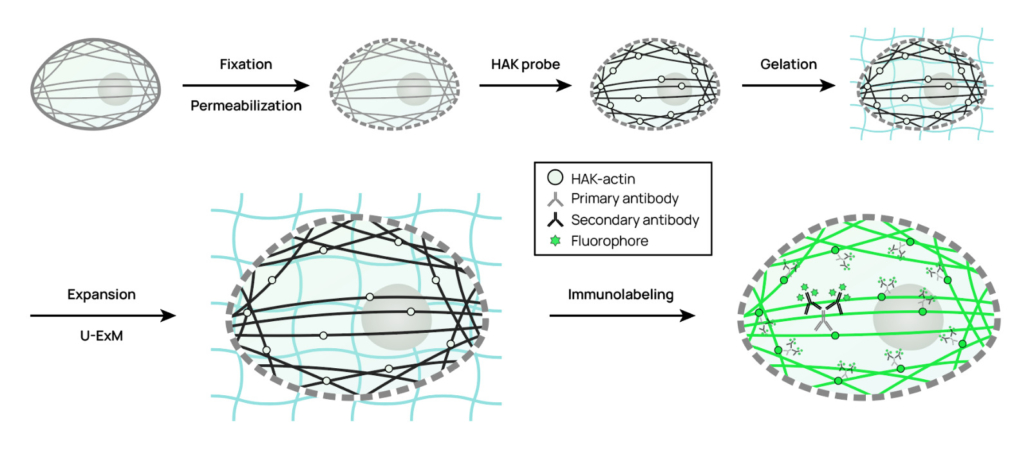
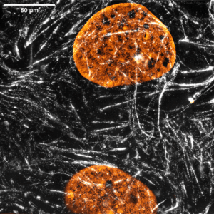
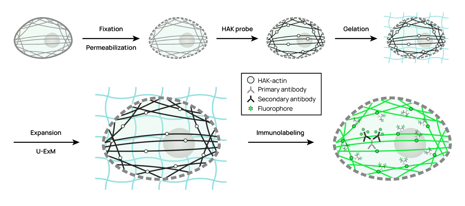
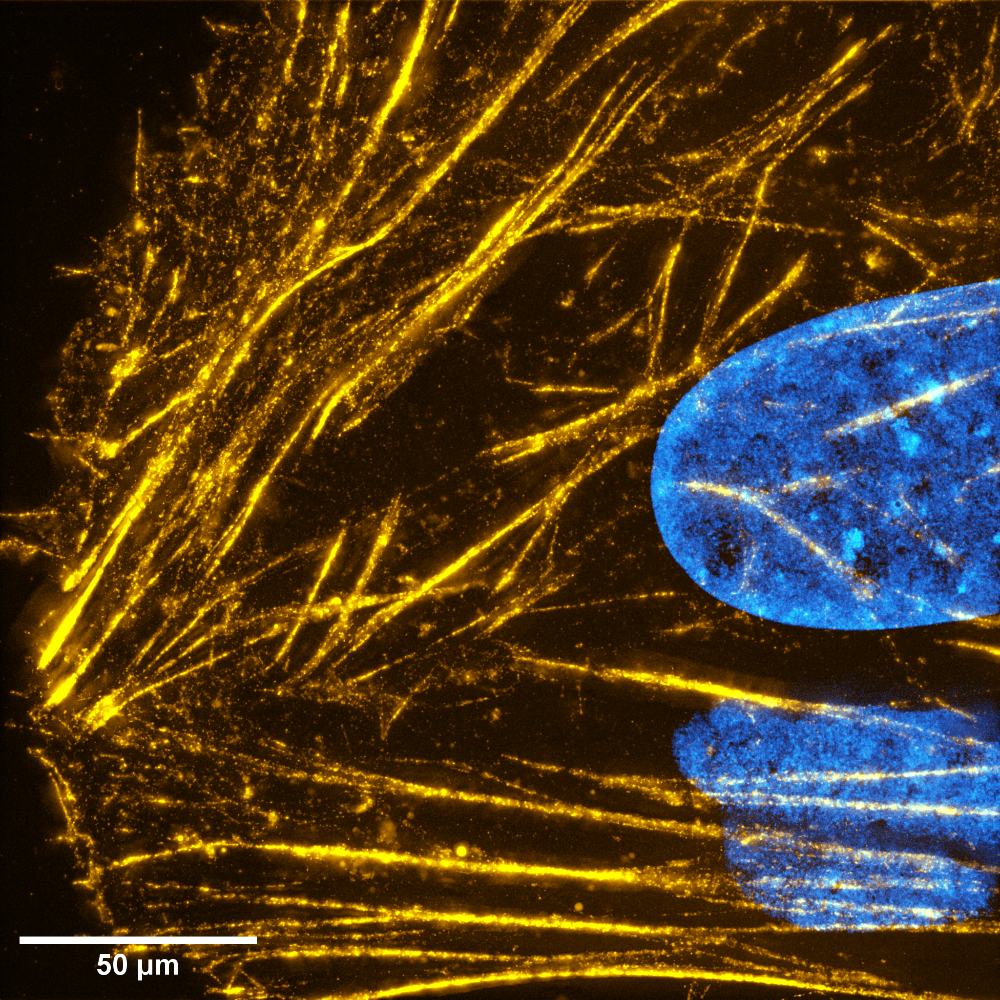
HAK-actin™ is a pan-species Expansion Microscopy (ExM) compatible probe for actin visualization using U-ExM based protocols. HAK-actin™ binds specifically to F-actin. Its design allows HAK-actin™ to be anchored into the gel and expand faithfully to the original actin structure after the swelling step. HAK-actin™ contains an HA tag epitope, it can therefore be detected post expansion using a pair of anti-HA and species specific secondary antibodies. It combines simplicity through a single protocol and versatility as the fluorophore label on the secondary antibody can be chosen freely. Finally, the primary + secondary antibody boosts fluorescent signal amplification, circumventing volumetric dilution of the fluorescent signal due to expansion.
Contains 1 tube of HAK-actin™ for 100 gels*.
How does HAK-actin™ work?
HAK-actin™ probe contains 3 key elements:
1. A pan-species, highly specific, high affinity F-actin ligand for a robust binding to polymerized actin (F-actin) only.
2. An anchoring moiety that specifically reacts with acrylamide and covalently links HAK-actin™ to the expansion gel.
3. The HA-tag epitope sequence for a strong and specific recognition by anti-HA antibodies, even after expansion.
HAK-actin™ key advantages
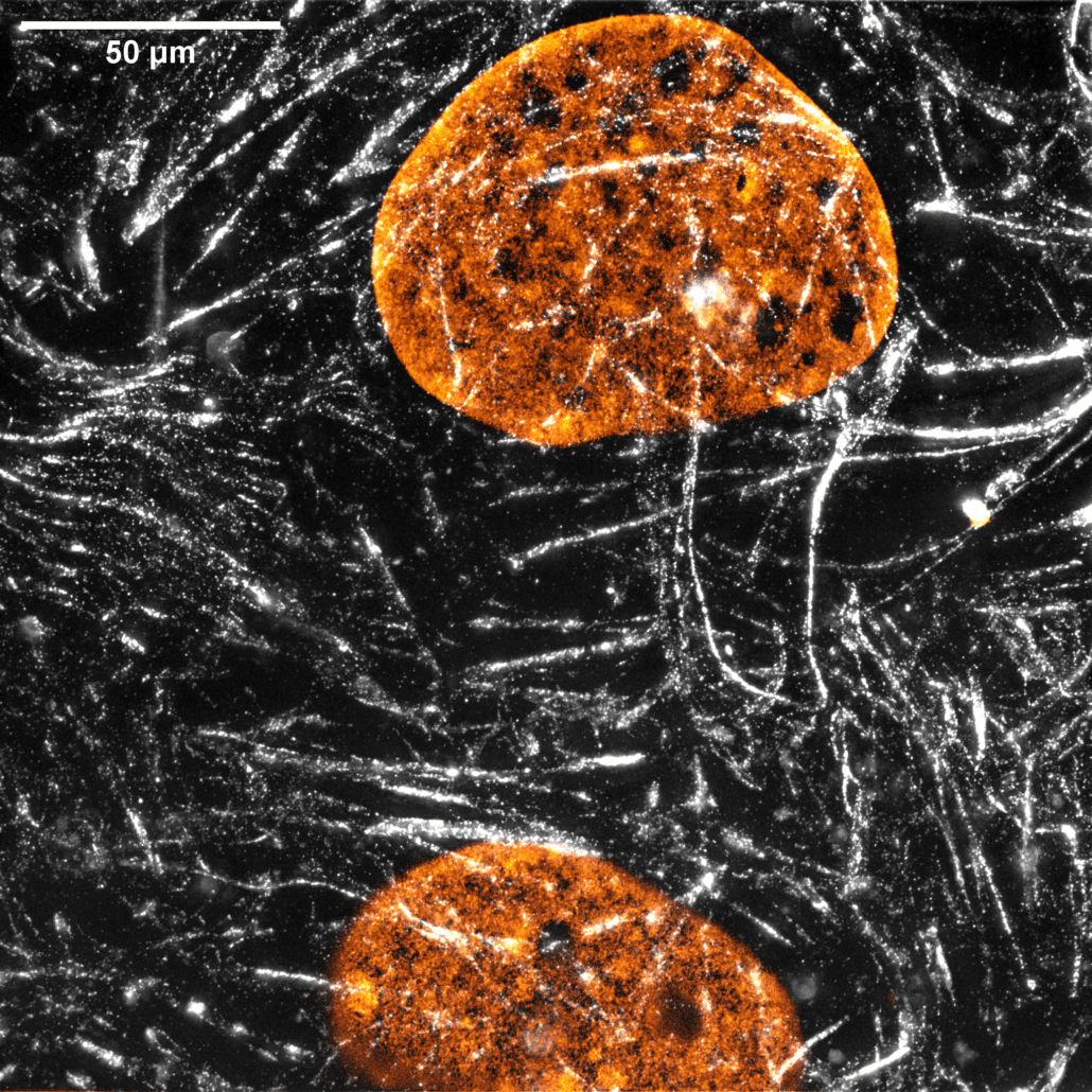
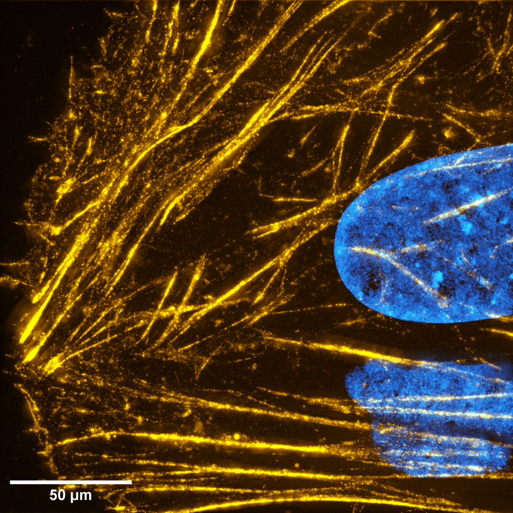
Stronger signal via post-expansion immunostaining. The HA tag allows post-expansion labeling with standard anti-HA antibodies, giving ~4× higher fluorescence than a directly labeled probe. You are free to choose any fluorophore on the secondary antibody.
Robust anchoring/retention in the gel. Four lysines promote efficient crosslinking to the U-ExM polymer, yielding bright, specific actin staining with low background.
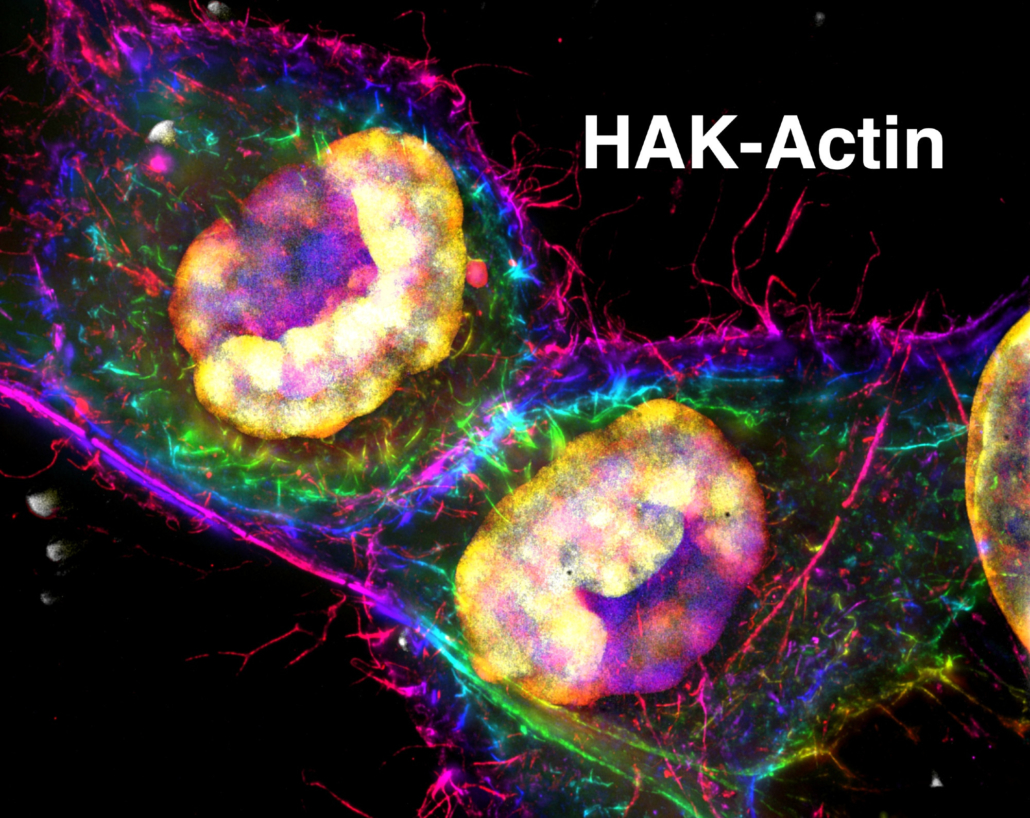
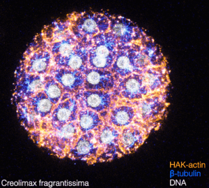
Wide protocol compatibility. Works with standard U-ExM, cryo-ExM, and iU-ExM (up to ~16× expansion). HAK-actin™ can be multiplexed with other targets (e.g., Microtubules).
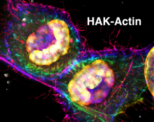
Broad applicability across models. Delivers uniform actin labeling in human cells (HeLa, U2OS), neurons (growth cones, dendritic spines), platelets, mouse retina (clearer signal in the ciliary bulge), and diverse microbial eukaryotes—including ichtiosporea cell-walled species where anti-actin or phalloidin may fail.
Overcomes key ExM limitations. By using post-labeling, it avoids the fluorophore dilution and color constraints of pre-labeling approaches, doesn’t require species-specific antibodies or actin genetic tagging, and simplifies a reproducible workflow.
No transfection or expression required. HAK-actin does not require genetic expression of tagged actin monomers. It labels endogenous actin.
Downloads:
References:
Manuscript describing the HAK-actin™: Mercey, O., et al: HAK-actin, U-ExM-compatible probe to image the actin cytoskeleton. BioRxiv, 2025.
U-ExM protocol article: https://doi.org/10.1016/bs.mcb.2020.05.006
iU-ExM protocol article: https://doi.org/10.1038/s41467-023-43582-8
Cryo-ExM protocol article: https://doi.org/10.1038/s41592-021-01356-4
*Based on the following conditions: 0.5 ml staining solution / staining experiments with 1x probe concentration. The number of staining experiments can be further increased by reducing volume or probe concentration.
HAK-actin™ probe is distributed by Spirochrome under an exclusive license from the University of Geneva, Switzerland. HAK-actin™ is a registered trademark of University of Geneva, Switzerland.

