
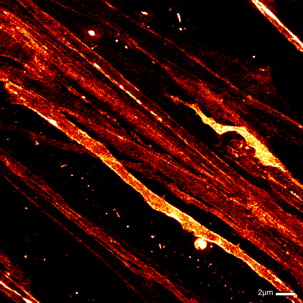
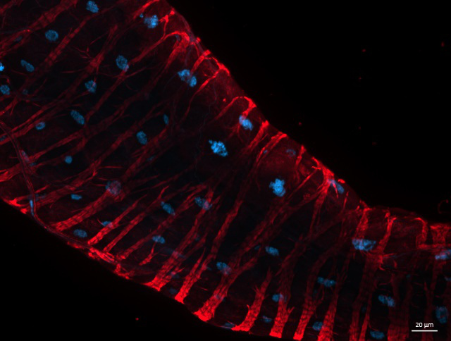
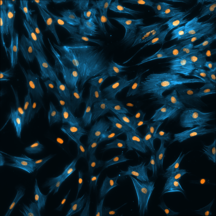
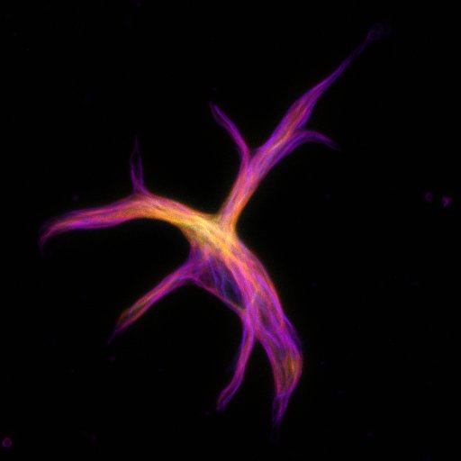
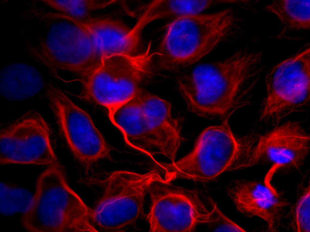
SiR-actin
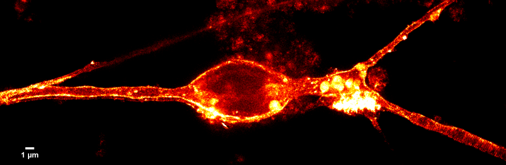
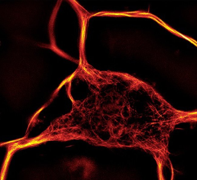
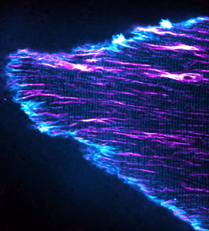
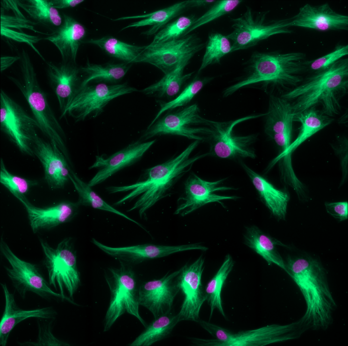
SiR-tubulin
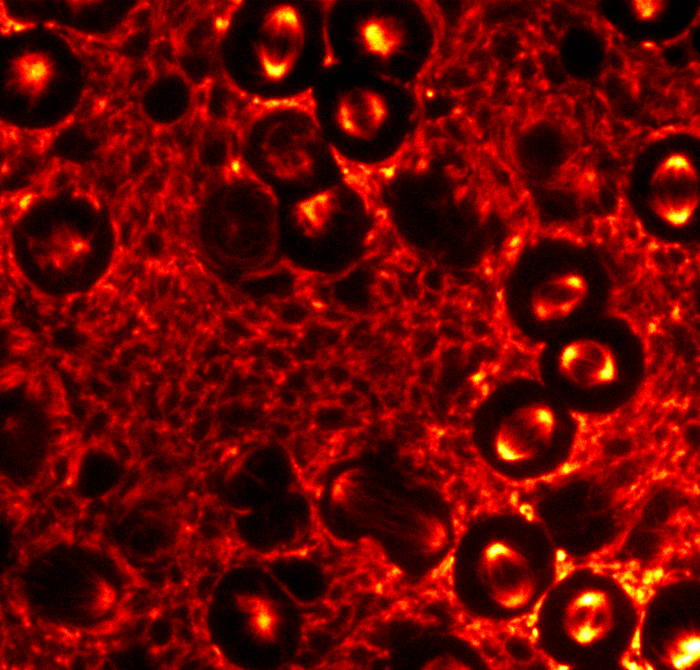
SiR-DNA
Movie Gallery
Movie of newborn mouse primary cardiac myocytes stained with SiR-actin. High speed (50fps) confocal imaging. Courtesy of Adam Kwiatkowski and Simon Watkins, department of cell biology and center for biologic imaging, University of Pittsburgh.
Movie of mouse oocyte from transgenic CAG::H2B-EGFP mouse stained with SiR-tubulin. Chromosomes (H2B) are pseudocolored in red, microtubules in green. Courtesy of Petr Solc, Institute of Animal Physiology and Genetics at Academy of Sciences of the Czech Republic, Brno.
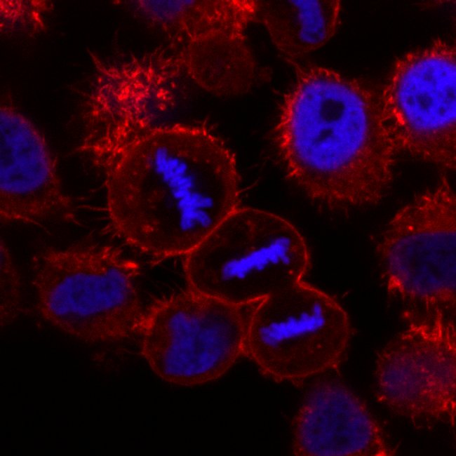
Movie of HeLa cell expressing mcherry-H2B (red) stained with SiR-tubulin (green). Data collected by confocal imaging. Courtesy of Daniel Gerlich and Claudia Blaukopf, Institute of Molecular Biotechnology, Vienna.
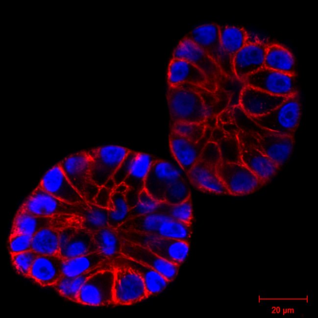
3D reconstruction from a z-stack of MCF10A cells spheroid stained with SiR-DNA. Courtesy of Christian Conrad and Katharina Jechow, Heidelberg.
Movie of Live HeLa cells stained with SiR-DNA. Data collected by confocal imaging. Courtesy of Daniel Gerlich and Claudia Blaukopf, Institute of Molecular Biotechnology, Vienna